


Background
Acute pulmonary embolism is rarely complicated by development of pulmonary hypertension (PH) with estimated incidence rate of 0,5-2%. It is almost impossible to determine the overall prevalence of chronic thromboembolic pulmonary hypertension (CTEPH) since not all of these patients have a history of acute pulmonary embolism. For patients after acute pulmonary embolism, diagnosis can be made after 3 months of effective anticoagulation, and remaining signs of PH. Final diagnosis can be made after confirming pre-capillary PH during right heart catheterization in patients with multiple chronic/organized occlusive thrombi/emboli in the elastic pulmonary arteries. Despite of severity, in some cases it is possible to cure patients with surgical pulmonary thromboendarterectomy (PTE).
Case presentation
We present a case of 41-years old caucasian male with CTEPH coexisting with B-cell chronic lymphocytic leukemia (B-CLL). First symptoms occurred during winter 2012 (mild dyspnea, fatigue), followed by enlargement of peripheral lymph nodes, night sweats, loss of weight (July 2012). In August 2012, patient developed severe dyspnea in class IV according to NYHA. During hospitalization, acute pulmonary embolism was discovered, as well as suspicion of non-Hodgkin lymphoma, and anticoagulation with oral vitamin K antagonists was introduced. Patient was admitted to the Centre of Rare Cardiovascular Diseases at John Paul II Hospital (CRCD) in November 2012, we decided to continue anticoagulation for at least 3 months, then to do comprehensive clinical re-assessment. Performed tests for congenital hypercoagulation disorders were all negative. Meanwhile the patient underwent hematological investigations, which confirmed diagnosis of B-CLL in stage of Rai I (Binet A). At admission to CRCD, patient was in functional class III, according to NYHA classification, in physical examination there were no peripheral edema, vesicular sounds over lungs were present, and he had palpable, enlarged lymphatic nodes. In cardiopulmonary exercise test he reached 3,5 METs, Vmax=7,9 ml/kg/min; AT 6,1 ml/kg/min. In a 6-minute walk test the patient walked for 400m distance, and his blood saturation dropped from 87% to 80%. Right heart catheterization results are showed in Table 1. The procedure confirmed elevated mean pressure in pulmonary artery (69 mmHg), low cardiac index and high pulmonary vascular resistance. An angiography of pulmonary arteries revealed multiple segmental and subsegmental perfusion defects (Figure 1 and 2).
Table 1. Right heart catheterization.
|
Pressure [mmHg] |
REST |
AFTER NO INHALATION |
|
RA |
6 |
6 |
|
PA |
106/46/64 |
103/41/59 |
|
RV |
99/-1/10 |
– |
|
PCWP |
9 |
4 |
|
LV |
– |
– |
|
AORTA |
118/67/79 |
111/60/73 |
|
Saturation [%] |
||
|
VCI |
49,2 |
– |
|
VCS |
38,0 |
– |
|
RV |
42,9 |
– |
|
PA |
44,7 |
57,2 |
|
AORTA |
83,2 |
87,9 |
|
Cardiac Output [l/min] |
3,21 |
4,05 |
|
Cardiac Index [l/min/m2] |
1,63 |
2,06 |
|
Qp/Qs |
1 |
– |
|
PVR [ARU] |
1370 |
1086 |
|
TPR [ARU] |
1595 |
1165 |
|
VSR [mmHg] |
1819 |
1441 |
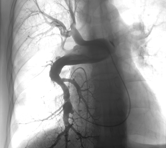
Figure 1. Right pulmonary artery angiography – AP aspect. Losses in perfusion in segmental and subsegmental arteries.
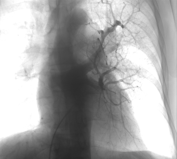
Figure 2. Left pulmonary artery angiography - AP aspect. Significant losses of perfusion are present in lower lobe.
